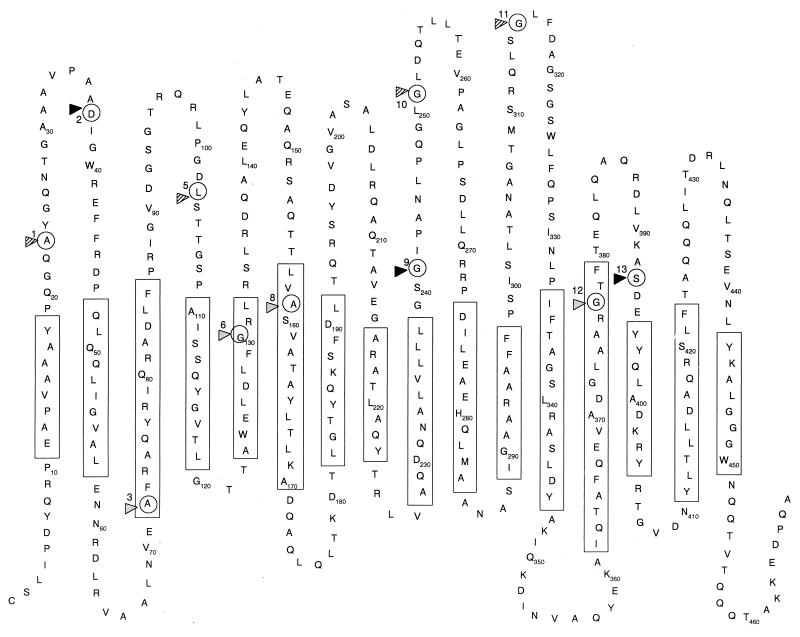
FIG. 2.
Predicted membrane topology model of the OprM monomer. Rectangles enclose the proposed transmembrane β strands. Circles indicate insertion sites ME1 to -3, -5, -6, and -8 to -13 (numbered correspondingly) for the malarial epitope. Permissive insertions are indicated by solid triangles, partially permissive ones are indicated by shaded triangles, and nonpermissive ones are indicated by gray triangles.

