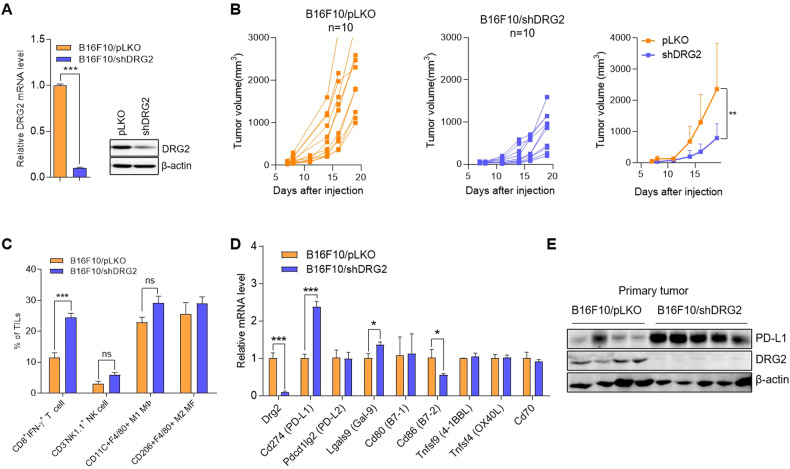
Fig. 1. DRG2 depletion in melanoma cells enhances PD-L1 expression in cancer cells but increases the proportion of tumor-infiltrating CD8 T cells and inhibits tumor growth.
A Confirmation of DRG2 depletion in B16F10/shDRG2 cells by qRT-PCR and western blot analysis. B Individual and mean tumor growth curves for mice s.c. injected with B16F10/pLKO or B16F10/shDRG2 cells. Data are pooled from two independent experiments (n = 10 per group). The data are expressed as means ± SD. Two-way ANOVA, **P < 0.01. C–E Tumor masses were collected 15 days after s.c. injection of melanoma cells. C FACS analysis for immune cells in TIICs of B16F10/pLKO and B16F10/shDRG2 tumors. Graph represents % of immune cells within TIICs. Values are the mean ± SD of two independent experiments (n = 3 per group per experiment). Student’s t test. ***P < 0.001. ns not significant. See also Supplementary Fig. S1. D qRT-PCR analysis for expression of immune checkpoint molecules in B16F10/pLKO and B16F10/shDRG2 tumors. Values are the mean ± SD of two independent experiments (n = 3 per group per experiment). Student’s t test. *P < 0.05; ***P < 0.001. E Western blot analysis for PD-L1 in B16F10/pLKO and B16F10/shDRG2 tumors.

