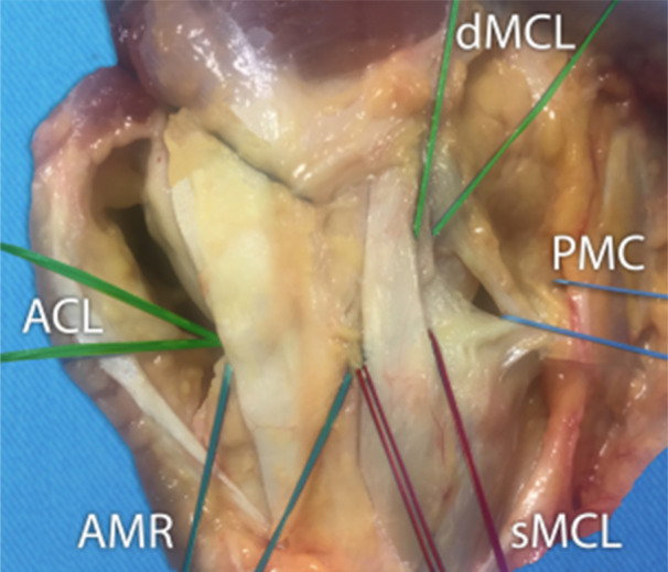
Figure 1.

Medial view of a right knee. The identification starts in the middle third as it represents the medial collateral ligament (MCL). This is divided into superficial (sMCL) and deep layers (dMCL). The anterior margin of this structure was clearly defined by palpation of longitudinal fibres but posteriorly the margins were less clear. At the posterior margin of the middle section, the longitudinal fibres blended with oblique fibres coming from the posterior third. This third part is composed of three layers: the facial layer (layer 1), the sMCL (layer 2) and the dMCL and the capsule (layer 3) according to Warren and Marshall's description. The MCL is attached on and around the femoral epicondyle. The anterior third named anteromedial retinaculum (AMR) is composed of fascia (layer 1) and capsule (layer 3) without substantial ligament structure linking the femur to the tibia. In the posterior third, layers 2 and 3 were not separated and formed the posterior medial capsule (PMC). The localised thicker band in the PMC is the posterior oblique ligament (POL). ACL, anterior cruciate ligament.
