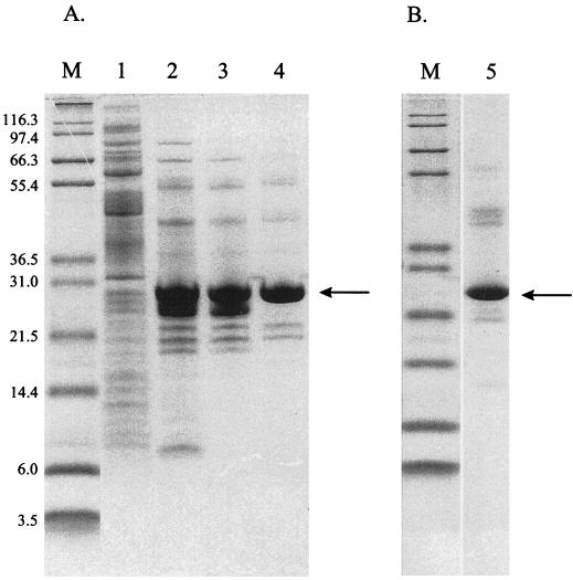
FIG. 2.
Peptidase E purification. (A) X. laevis peptidase E was purified from soluble cell extracts of strain TN5415 as described in Materials and Methods. Samples from each step in the purification were analyzed by SDS-PAGE and loaded as follows: crude cell extract (lane 1), pooled Q Sepharose fractions (lane 2), pooled Superose fractions (lane 3), and pooled DEAE fractions (lane 4). (B) Serovar Typhimurium peptidase E, which was purified as described in Materials and Methods, was subjected to SDS-PAGE and loaded in lane 5. Molecular mass markers were loaded in lanes labeled “M,” and their masses (in kilodaltons) are noted to the left of the gels. The band on each gel corresponding to peptidase E is indicated with an arrow.

