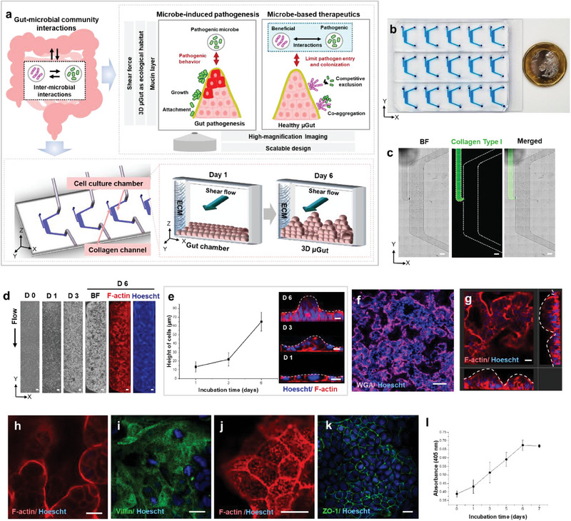
Figure 1.

The GMoC showcasing a dynamic 3D µGut‐microbes interface, where the shear‐induced 3D µGut effectively mimics the key structures and functions of the human intestine. a) The GMoC consisting of µGut chamber consisting of the cell culture chamber and the collagen channel, features a dynamic gut‐microbiome interface. The chip provides shear flow and the ECM‐supported self‐structured 3D µGut capable of producing mucin, supporting microbial attachment and growth over an extended period. The chip offers compatibility with high‐magnification imaging to visualize a single microbe, distinguish different bacterial species and their inter‐microbial interaction as well as the influence of a microbial community on the µGut. Our GMoC allows for dissecting causal and mechanistic roles of gut microbes for microbe‐induced pathogenesis (Figures 3 and 4) and microbe‐based therapeutics (Figures 5 and 6). b) The GMoC featuring high scalability and integrability to high‐magnification imaging. c) Brightfield and fluorescence images of the chip showcasing the collagen (FITC) channel and the culture chamber (Scale bar, 500 µm). d) Time‐lapse images of the Caco‐2 derived µGut formation in the GMoC over 6 days. Fluorescence images of the µGut on Day 6 show uniform cell density across the length of the chip (Scale bar, 200 µm). e) Cell height measurement and cross‐section images of the µGut over 6 days showing morphogenesis of a villus‐like structure. f) Confocal immunofluorescence top‐view image of the 3D stratified µGut epithelium (Scale bar, 100 µm) consisting of g) continuous crypt‐villus units. h) Top view of villi‐like structures, and i) brush border covered with j) microvilli in the µGut. k) Expression of tight junction (ZO‐1) indicating gut barrier formation. l) Steady increase of aminopeptidase activity of the μGut during the self‐morphing process. All scale bars represent 20 µm unless otherwise indicated.
