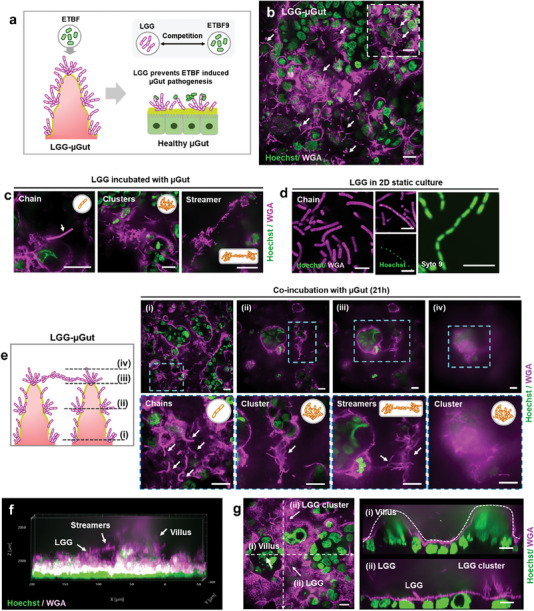
Figure 5.

Establishment of the dynamic probiotic‐enriched µGut (LGG‐µGut), showcasing the formation of distinctive LGG structures. a) A schematic illustrating pre‐treatment of the µGut with beneficial LGG (LGG‐ µGut) resisting ETBF colonization and growth by inter‐microbial competition, preserving the healthy state of µGut. b) Top view of the LGG‐µGut showing the abundance of LGG (Pink strands). The high background from the epithelial cells is due to the gram stain, WGA, staining the mucin on the epithelial cells. The inset figure shows protruding LGG strands from the mucin layer on the µGut epithelium. c) Different LGG structures formed in the LGG‐µGut. d) Chain forming LGG in MRS agar media (Scale bar, 5 µm). e) Confocal Z‐stack images of the LGG‐µGut from the bottom i) to top planes iv) of the crypt‐villus axis showcasing various LGG structures. f) 3D visualization of the LGG‐ µGut showing the abundant presence of LGG covering the villus‐crypt axis. g) Cross‐section images of the LGG‐ µGut showcasing i) undisrupted villi‐like structure covered with ii) LGG chains and clusters. All scale bars represent 20 µm unless otherwise indicated.
