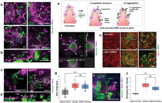
Figure 6.

LGG‐µGut exhibit resistance against ETBF colonization through effective competitive mechanisms, protecting the µGut from ETBF‐induced disruption. a) Diminished ETBF9 colonization in the LGG‐µGut evidenced by the presence ofi) only a few ETBF9 and ii) small‐sized ETBF9 colonies on the LGG‐colonized surface. b) A cross‐section image of an ETBF9 colony in the LGG‐µGut showing the attachment of ETBF9 on LGG. c) Top view and d) cross‐section of ETBF9 co‐aggregating with LGG chains or LGG biofilms. e) A schematic diagram of the two direct colonization resistance mechanisms of the LGG‐µGut against ETBF. i) LGG inhibits ETBF adhesion or proliferation by competitive exclusion or (ii) LGG co‐aggregate with ETBF9, preventing ETBF9 access to the µGut surface. f,g) Cross‐section and height measurement of villi‐like structures in the LGG‐µGut after ETBF9 colonization. h,i) Immunofluorescence confocal images of ZO‐1 and j) normalized ZO‐1 density in the LGG‐µGut after ETBF9 colonization. All scale bars represent 20 µm unless otherwise indicated.
