
Figure 3.
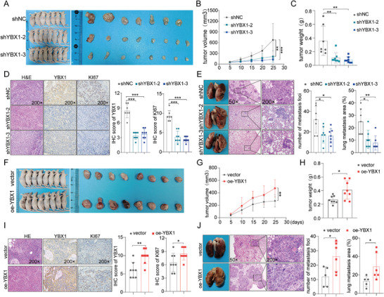
YBX1 facilitates xenograft tumor growth and lung metastasis of ESCC in vivo. A) Morphological pictures of the decreased subcutaneous xenograft tumor formation in mice injected with YBX1‐depleted KYSE150 cells. B,C) Tumor volume growth curves and final tumor weight were measured and quantified. D) Representative H&E and IHC images (left) of the tumor described. IHC staining data of YBX1 and KI67 of the mice in each group was quantified (right). E) Decreased tumor metastasis in mouse lungs with YBX1‐depleted KYSE150 cells, as determined by tail‐vein injection metastasis assays. Images of the mouse lungs with metastatic nodules and corresponding H&E staining of the metastatic tumors were presented and quantified for analysis. F) Morphological images of orthotopic‐xenograft mouse models implanted with YBX1‐overexpression in KYSE30 cells. G,H) Tumor growth and tumor weight were measured and quantified. I) H&E and IHC staining of the tumor (left). IHC staining data of YBX1 and KI67 of the mice in each group were quantified (right). J) Representative images of the overall observation of lungs with metastatic nodules and their corresponding H&E images (left). Lung metastasis nodules were further quantified (right). * p < 0.05, ** p < 0.01, and *** p < 0.001.