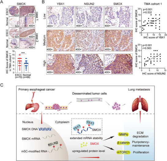
Figure 8.

SMOX expression was positively correlated with YBX1 and NSUN2 in ESCC. A) Representative IHC staining images of SMOX in ESCC tissues and adjacent normal tissues (up). IHC staining data of SMOX was quantified (down). B) Representative IHC images (×400 magnification) of YBX1, NSUN2, and SMOX in ESCC tissues with low or high expression (left). Scale bars, 50 µm. Spearman's correlation analysis was performed (right). C) Proposed model illustrating the functional role of YBX1/m5C‐SMOX axis in facilitating the progression of ESCC.
