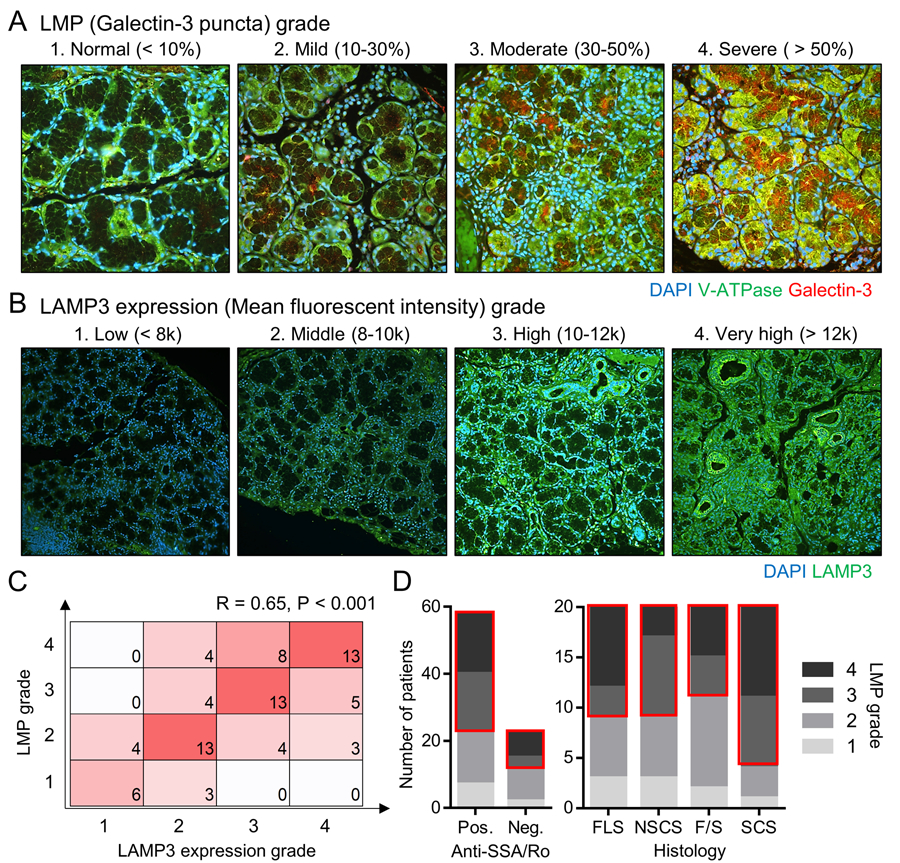
Figure 2. LMP is correlated with LAMP3 expression in salivary glands of SjD patients.

(A, B) Labial minor salivary gland sections from SjD patients (n = 80) were stained for (A) V-ATPase (green) and galectin-3 (red) (original magnification: 40x) or for (B) LAMP3 (green) (original magnification: 20x). Representative immunofluorescent image in each LMP and LAMP3 expression grade are shown. (C) Matrix shows the number of patients having indicated combination of LMP and LAMP3 expression grades. (D) Bar charts show the number of patients having indicated LMP grade based on positivity of serum anti-SSA/Ro antibodies or salivary gland histology. FLS, focal lymphocytic sialadenitis; NSCS, non-specific chronic sialadenitis; SCS, sclerosing chronic sialadenitis; F/S, combination of FLS and SCS features.
