
Figure 4. Caspase-8 is degraded through chaperone-mediated autophagy.
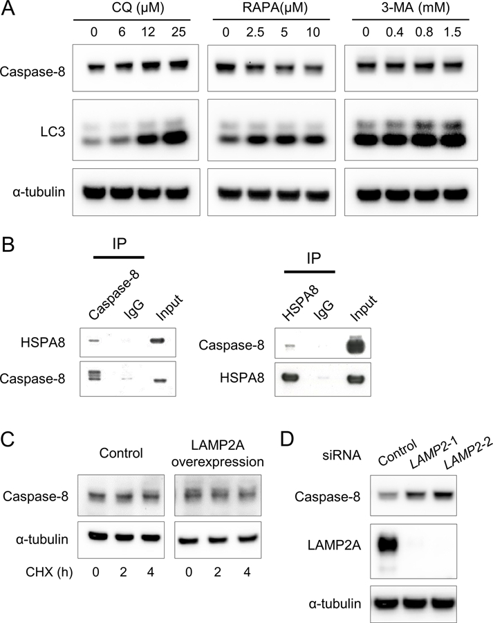
(A) Western blots showing caspase-8, LC3, and α-tubulin (internal control) expressions in A253 cells treated with chloroquine (CQ), rapamycin (RAPA), or 3-methyladenine (3-MA) at indicated concentration for 24 hours. (B) Western blots showing HSPA8 and caspase-8 expressions immunoprecipitated from A253 cell lysates using anti-caspase-8 antibodies or anti- HSPA8 antibodies, respectively (C) Western blots showing caspase-8 and α-tubulin expressions in A253 cells treated with cycloheximide (CHX) at 50 μg/mL for the indicated hours 48 hours after transfection with LAMP2A expression plasmids. (D) Western blots showing caspase-8, LAMP2A, and α-tubulin expressions in A253 cells treated with negative control or two types of LAMP2 siRNA for 48 hours.