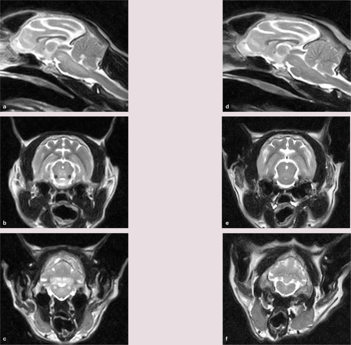
Figure 2.
(a) Midline sagittal section showing intra-axial regions of hyperintensity to grey matter in the mesencephalon and pons. (b and c) Transverse images at the level of the rostral colliculus (b) and caudal cerebellar peduncles (c). Bilaterally symmetrical hyperintense to grey matter lesions can be identified affecting the mesencephalic tegmentum (adjacent to the black asterisks) and white matter lesions affecting the caudal cerebellar peduncles (adjacent to the white asterisks). (d f) Magnetic resonance images corresponding to images (a c) following 8 weeks of cobalamin supplementation. The hyperintense lesions have completely resolved

