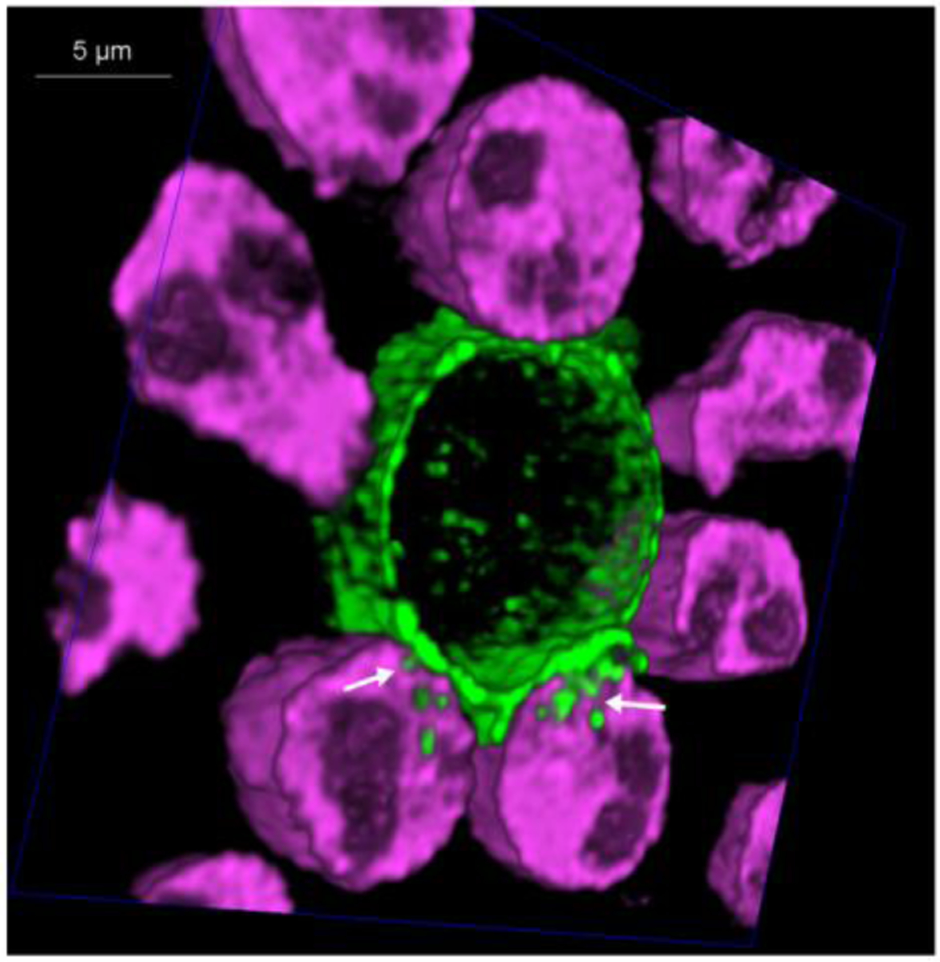
Figure 2:

Human neutrophils kill Trichomonas vaginalis using trogocytosis: Primary human neutrophils were labelled with Cell Tracker Deep Red (magenta) and the Trichomonas vaginalis surface was biotinylated and labelled with streptavidin-488 (green). Neutrophils and parasites were allowed to interact at a ratio of 10 neutrophils: 1 parasite in the presence of 10% human AB serum at 370C. HyVolution, super-resolution live confocal imaging (Leica Microsystems) was used to take 60 cross-sections of the cells interacting in real-time. A cross-section of a 3 –dimensional image is shown, revealing neutrophil trogocytosis (nibbling) of the parasite. Location of “nibbling” in action is noted with white arrows. Complete data and methods are published [70].
