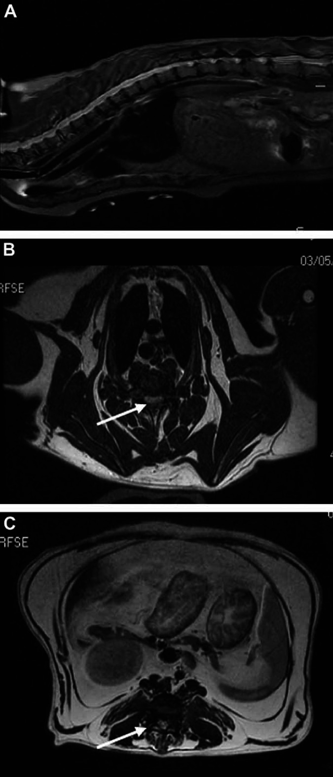
Fig 1.
MRI of multifocal feline IVDD. (A) MRI of sagittal view at the level of the midline. (T2 SAG FS): multiples areas of disc protrusion and degeneration throughout the thoracic and cranial lumbar spine were noted. Spinal cord compression at intervertebral spaces from T2 to T6 (n=4) and from L2 to L5 (n=3) was more pronounced. (B) Transverse MRI (AX T2 FRFSE) of the most severe disc protrusion by centrally protruding intervertebral disc at T3–T4 intervertebral disc space. (C) Transverse MRI of the lumbar space disc protrusions at the L3–L4 intervertebral disc space (AX T2 FRFSE) and moderate severe compression by centrally protruding intervertebral disc from L3 to L4 was remarkable.

