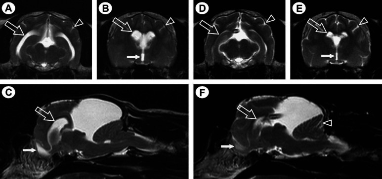
Fig 1.
T2-weighted transverse (A, B, D and E) and para-sagittal (C and F) MR images, demonstrating a large hyperintense, cystic lesion dorsal to the colliculi (C and E). Images A–C are prior to cystoperitoneal shunt placement and images D–F are post cystoperitoneal shunt placement. The MR imaging appearance is consistent with a quadrigeminal cyst compressing the cerebellum and brain stem ventrally and the cerebral hemispheres rostrally. Evidence of obstructive hydrocephalus resulting in increased intracranial pressure is evident as enlargement of the lateral (open arrows in A–C) and third (filled arrow in B) ventricles, dilation of the olfactory bulb cavity (filled arrow in C), attenuation of the hyperintense CSF signal within the cerebral sulci (arrowhead in A and B) and between the folia of the cerebellum and displacement of the caudal aspect of the cerebellum into the cervical spinal canal. Resolution of the features suggestive of secondary obstructive hydrocephalus is evident following placement of a cystoperitoneal shunt (D–F), with the shunt-tip visible in images D and F.

