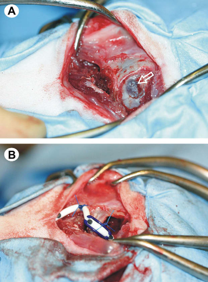
Fig 2.
Intra-operative images, demonstrating cystoperitoneal shunt placement. In both images, rostral is to the right and caudal to the left. The apposed dura and cyst wall are visible within the craniectomy defect (open arrow in A). Successful placement of the cystoperitoneal shunt, with fixation of the shunt tubing to the periosteum of the occipital bone (B).

