Figure 2.

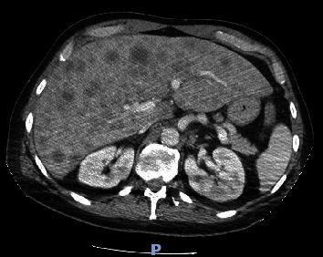
Axial section obtained from computed tomography of the abdomen and pelvis shows numerous hypovascular liver masses measuring up to 4.8 cm.

Axial section obtained from computed tomography of the abdomen and pelvis shows numerous hypovascular liver masses measuring up to 4.8 cm.