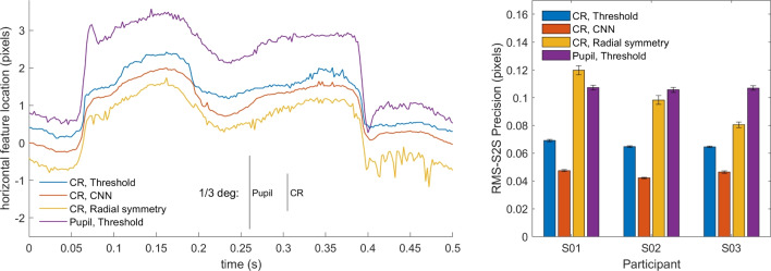
Fig. 7.
Real eye CR and pupil center signals of dataset one. Left: representative segment of pupil and CR center signals from S03 in camera pixels. For the CR center, the signals produced by three different CR center localization methods is shown. The signals contain two small saccades and have been vertically offset for clarity. RMS precision for the shown segments are 0.081 px for the threshold signal, 0.061 px for CNN, and 0.096 px for radial symmetry and 0.121 px for the pupil signal. Further, an RMS precision comparison (right panel) between the three methods and the pupil signal on all data of three participants is shown. Error bars depict standard error of the mean

