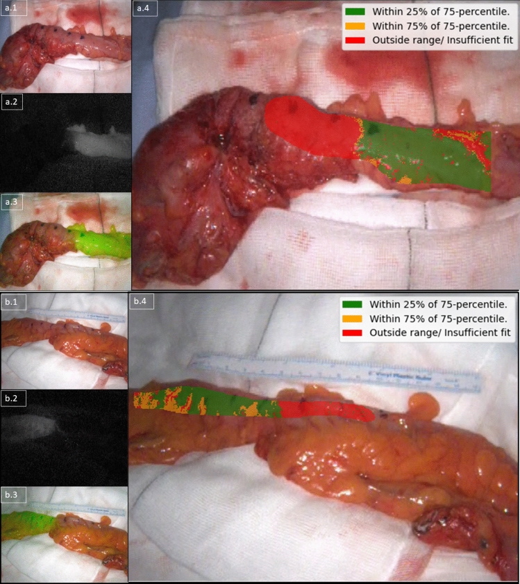
Fig. 4.
A composite image illustrating an augmented reality per pixel transection recommendation using Formula 1 for a sigmoid resection for diverticular disease on top (a, proximal on the right) and for T1N1 adenocarcinoma below (b, proximal on the left). The panel on the top left (a.1 and b.1) shows the white light view, the one in the middle left (a.2 and b.2) shows the raw NIR feed, and the ones on the bottom left (a.3 and b.3) show an overlay view (composed of the NIR feed overlaid on the white light feed). These three images are unchanged from the display of the camera system. The main large panels on the right (a.4 and b.5) show the computed recommendation augmented on the white light view. Computational recommendation (legend top right of a.4 and b.4) with green indicating sufficient, orange borderline and red insufficient perfusion (see supplementary material)

