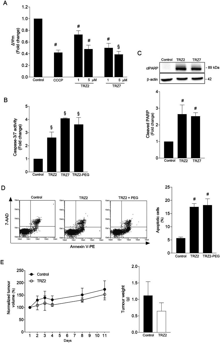
Fig. 3. Ru-TRZ hybrids induce mitochondrial depolarization and apoptotic cell death.
Ru-TRZ effects were determined in HT29 cells and HT29 xenograft tumor-bearing mice. A Mitochondria transmembrane potential (ΔΨm) was evaluated by JC-1 staining of HT29 cells exposed to TRZ2 and TRZ7 for 2 h or CCCP (200 µM) for 4 h (positive control). Data presented correspond to the ratio of fluorescence emitted at 590 and 530 nm. B Caspase-3/7 activity was determined in HT29 cells exposed to 10 μM of indicated Ru-TRZ compounds for 24 h, using the Caspase-Glo 3/7 assay. C Representative immunoblot of cleaved PARP-1 (clPARP) in whole-cell extracts from HT29 cells exposed to indicated Ru-TRZ compounds for 24 h and respective quantification normalized to endogenous β-actin. D Apoptotic cell death was analyzed by flow cytometry using the Guava Nexin assay, following incubation of HT29 cells with indicated Ru-TRZ compounds (10 µM), or vehicle, for 24 h. Representative flow cytometry plots (left) and respective quantification (right). Results are expressed as mean ± SEM from at least three independent experiments. E Normalized tumor volume evolution of TRZ2- and vehicle-treated HT29 xenograft tumor-bearing mice. Line graph represents tumor volume normalized to baseline at day 1 (left). Bar graph represents tumor weight after 20 days of treatment with TRZ2 or vehicle control (right). Results are expressed as mean ± SEM of the tumor volume or weight of at least three animals in each group. DMSO is the vehicle control. #p < 0.05 and §p < 0.001 from control cells; *p < 0.05 and $p < 0.001 from Ru-TRZ-treated cells.

