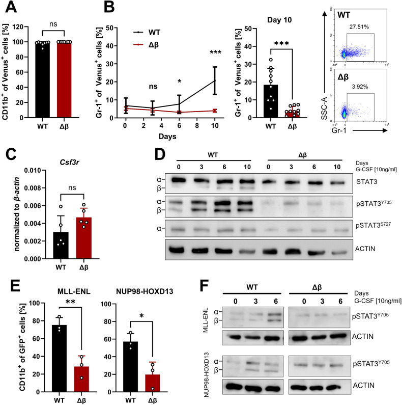
Fig. 2. Leukemia cells lacking STAT3β exhibit reduced differentiation response to G-CSF.
A Flow cytometry quantification of CD11b+ cells after 10 days cultured with 10 ng/ml G-CSF. (n = 8, 3 independent experiments, 2–3 cell lines). B Quantification of Gr-1+ cells after 10 days of G-CSF stimulation via flow cytometry and representative dot plots from day 10 (n = 10, 4 independent experiments, 2–3 cell lines). C RT-qPCR analysis of G-CSFR (Csf3r) expression in suspension culture (n = 5, 3 independent experiments, 2 cell lines). D Western blot of G-CSF stimulated samples with the indicated antibodies and time points. E Immunophenotyping of MLL-ENL and NUP98-HODX13 transformed FLCs after 10 days G-CSF via flow cytometry (n = 3, independent experiments). F Western blot of G-CSF stimulated samples (MLL-ENL and NUP98-HOXD13 transformed FLCs) with the indicated antibodies and time points. Statistical analysis was performed using Student’s t test and indicated as p ≤ 0.05:*, ≤0.01:**, ≤0.001:***. Error bars represent means ± SD.

