Figure 1.
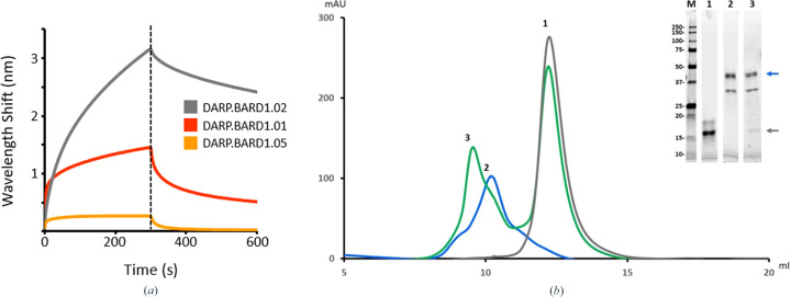
Verification of DARPin–BARD1 binding by biolayer interferometry (BLI) and analytical size-exclusion chromatography (AnSEC). (a) Putative anti-BARD1 DARPin molecules were screened for BARD1 binding by loading His-tagged DARPins onto NTA Biosensors and incubating with BARD1 for 5 min and then with analyte-free buffer for 5 min. The large wavelength shifts for DARP.BARD1.01 and DARP.BARD1.02 are indicative of strong antigen binding. (b) The size-exclusion profile shows that a DARPin–BARD1 mixture (peak 3) has an altered retention time relative to BARD1 (peak 2) or the anti-BARD1 DARPin (DARP.BARD1.02; peak 1) alone. Inset: SDS–PAGE analysis of the peaks from AnSEC purification. Lane M, broad-range molecular-weight marker; lanes 1, 2 and 3 correspond to peaks 1, 2 and 3, respectively. The blue and gray arrows indicate the positions of BARD1 and DARP.BARD1.02 DARPin, respectively.

