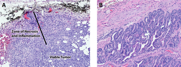
Figure 4.

Representative H&E images of prostate and prostate tumor from dog #3. A. Prostate tumor after PDT treatment. The black dye is a surface dye used to localize the region of PDT laser treatment. The tumor near the surface had necrosis, hemorrhage, and secondary inflammation. The deeper tumor was viable. B. Normal adjacent prostate with benign prostatic hyperplasia and PDT therapy. There was no evidence of necrosis as a result of the PDT therapy.
