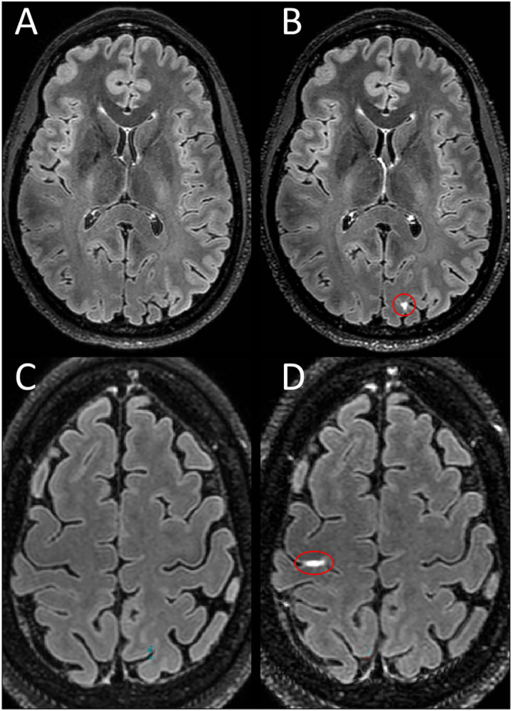
Fig 4. LME in healthy controls.

Shown are axial Gd+ Delayed 7T FLAIR images with LME highlighted by red ovals. Examples of LME in healthy controls as seen in a 40-year-old woman (B) and a 47-year-old woman (D) with no known neurological conditions. No leptomeningeal hyperintensity is seen in the same location on pre-contrast images (A,C).
