Figure 7.

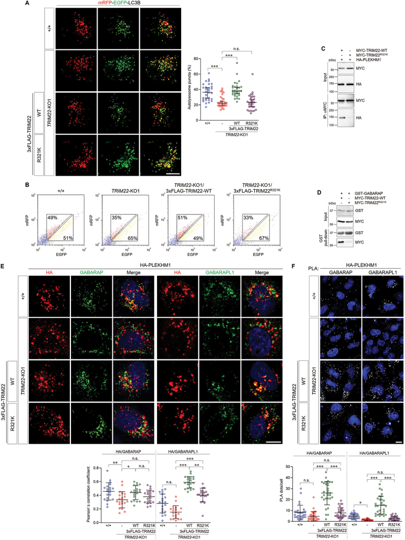
The R321K variant inhibits the role of TRIM22 in autophagosome-lysosome fusion. (A,B) mRFP-EGFP-LC3B was stably expressed in wild-type HeLa cells, TRIM22-KO1 cells, and TRIM22-KO1 cells expressing 3×FLAG-TRIM22-WT or R321K. (A) The cells with fluorescent LC3B puncta were analyzed. Puncta with mRFP+/EGFP+ indicate autophagosomes, and puncta with mRFP+/EGFP− indicate autolysosomes. Graphs show median with 95% confidence interval of more than 30 cells per group. (B) Cells were analyzed using flow cytometry. The intensities of mRFP and EGFP signals from more than 2,000 cells were analyzed, and the percentages of cells in corresponding gates are indicated. (C) HEK293T cells were transfected as indicated, and lysates were immunoprecipitated with antibodies against MYC. Precipitates were then immunoblotted as shown. (D) HEK293T cells were transfected as indicated, and lysates were incubated with GST-GABARAP proteins and precipitated with glutathione sepharose 4B beads. Precipitates were then immunoblotted as shown. (E,F) Wild-type HeLa cells, TRIM22-KO1 cells, and TRIM22-KO1 cells expressing 3×FLAG-TRIM22-WT or R321K were transfected with HA-PLEKHM1. (E) The cells were immunostained as indicated. Colocalization of HA-PLEKHM1 with GABARAP (left) or GABARAPL1 (right) was analyzed via calculation of Pearson’s correlation coefficient. Graphs show median with 95% confidence interval of more than 20 cells per group. (F) Cells were subjected to PLA with anti-HA antibodies along with antibodies against GABARAP (left) or GABARAPL1 (right). The number of PLA dots was quantified. Graphs show median with 95% confidence interval of more than 30 cells per group. *p < 0.05; **p < 0.01; *** p < 0.001; n.s., not significant. Scale bars: 10 μm.
