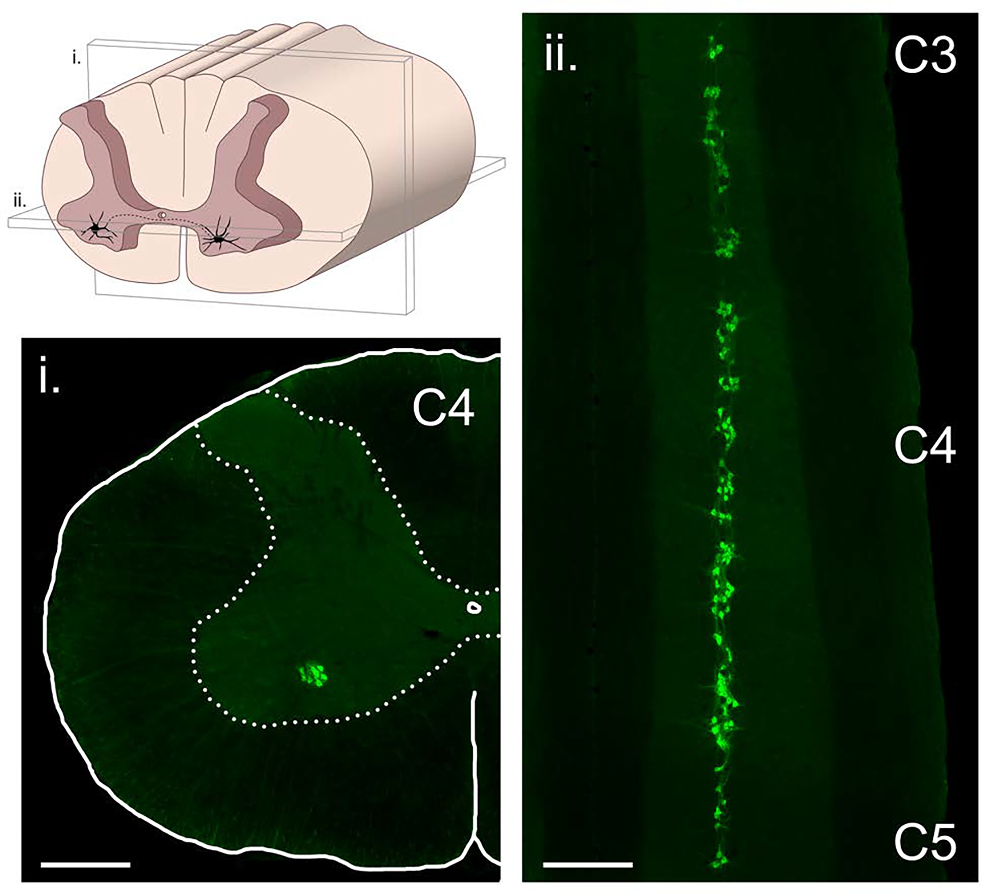
Figure 3. The phrenic motoneuron pool.

The column of phrenic motoneurons extends from approximately C3 to C5. The drawing in the upper left panel illustrates the location of phrenic motoneurons in the anterior horn, and also the plane of section that the two histological images were taken from. The histological images were obtained using a retrograde neuronal tracer (cholera toxin, ß-subunit) applied intrapleurally to the diaphragm muscle in an adult rat model. Scale bars indicate 500 μm.
