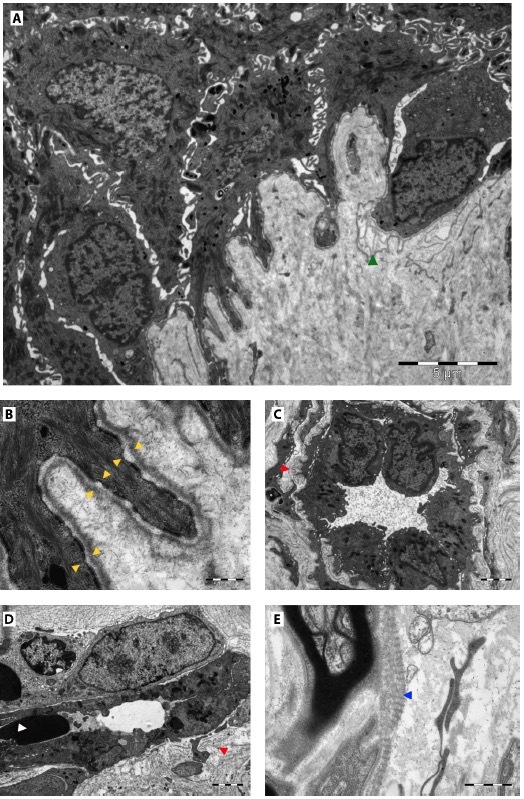
Figure 2.

Electron microscopy reveals focally disrupted hemidesmosomal internal/external plates in the basal layer (yellow arrowheads) with focal multiplication of basal lamina (green arrowhead) (A, B), mild dilation of superficial dermal vessels exhibiting multi-laminated and focally deformed basal laminas (suggestive for cutaneous collagenous vasculopathy) (red arrowheads) (C), and increased activity of endothelial cells. Perivascular lymphocytes and single extravasated erythrocytes can be noted (white arrowhead) (D). Focal abnormally banded (long-spaced) collagen (Luse-like bodies) in the vicinity of sensory receptors is suggestive for the diagnosis of CCV (blue arrowhead) (E).
