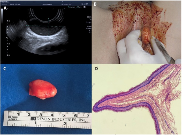
Figure 1.

(A) Perineal ultrasound demonstrated a unicameral well-demarcated oval-shaped cyst. (B) Intraoperative view of mass in the left labia majora. (C) The cyst was excised intact. (D) Histopathology: the cystic wall is serpiginous, lined by thin squamous epithelium with an outer corrugated cuticle and minimal granular layer (H&E, x20).
