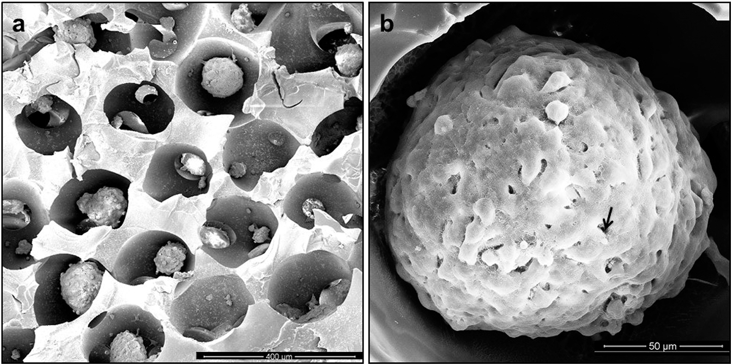
Figure 1.

SEM images of (a) dehydrated hydrogel ICC scaffolds cultured with cellular spheroids. Shape and pore diameter were shrunk during the dehydration process of SEM preparation. (b) SEM images of a mature spheroid in an ICC scaffold cultured for 5 days. Maturation of the spheroid is accompanied by formation of a layer of extracellular matrix on its surface, and individual cells become difficult to distinguish in the electron microscopy images. Scale bars: 400 μm (a) and 50 μm (b).
