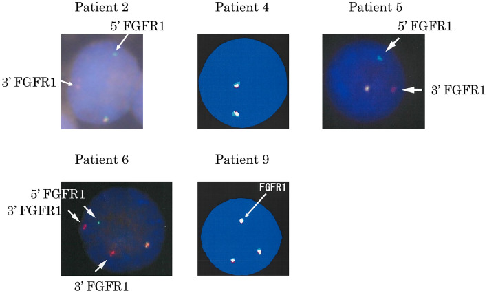
Fig. 1.
FGFR1 cleavage FISH on interphase nuclei. 5ʹ FGFR1 FISH probe was labeled green and 3ʹ FGFR1 FISH probe was labeled red. Fusion signal (red/green or yellow, white) indicates unaffected FGFR1. Splitting of the fusion signal (separated red or green signal) marks the rearrangement involving the FGFR1 locus. A split FGFR1 signal was detected in patient 2, patient 5, and patient 6. Amplification of the FGFR1 signal was detected in case 9

