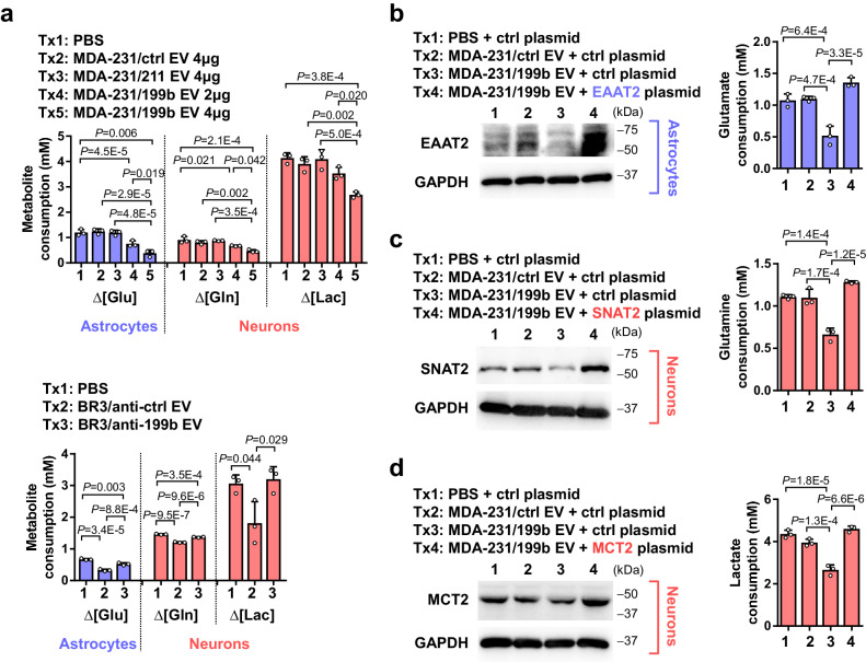
Fig. 4. EV miR-199b suppresses metabolite influxes in astrocytes and neurons.
a Astrocytes and neurons were treated with indicated EVs for 48 h and then changed to the media described in Methods. Net consumption of each metabolite was determined by calculating the difference of metabolite levels in the medium at the start and end points. To determine glutamate consumption, a starting level of 3 mM glutamate was used and the remaining glutamate in the culture medium of astrocytes was measured 24 h later (n = 3 biological replicates). To determine glutamine consumption, a starting level of 2 mM glutamine was used and the remaining glutamine in the culture medium of neurons was measured 48 h later (n = 3 biological replicates). To determine lactate consumption, a starting level of 25 mM lactate was used and the remaining lactate in the culture medium of neurons was measured 12 h later (n = 3 biological replicates). b–d Astrocytes (b) or neurons (c, d) were transfected with an overexpressing plasmid encoding EAAT2, SNAT2, or MCT2, or with the vector as a control, before treatment with EVs. Metabolite assays were carried out as in a (n = 3 biological replicates). In bar graphs, values are shown as mean ± SD. One-way ANOVA followed by Tukey’s multiple comparison test was used in a–d. P values are indicated. Source data are provided as a Source Data file.

