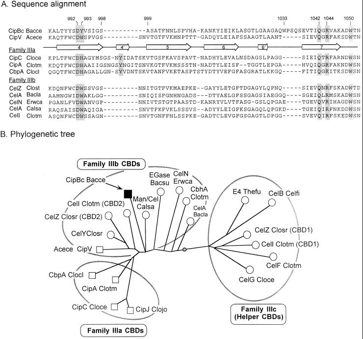
FIG. 5.
Relationship of the CipBc CBD to other scaffoldin and nonscaffoldin family III CBDs. (A) Sequence alignment of portions of selected family III CBDs, encompassing β-strands 4 through 7 (enumerated arrows). The CipBc CBD and the recently sequenced Acetivibrio CipV CBD (7) are compared to other known scaffoldin CBDs from family IIIa and nonscaffoldin family IIIb CBDs. Shaded residues indicate proposed cellulose-binding residues (39), and numbers refer to presumed positions on the mature CipBc protein. Dashes indicate gaps. (B) Phylogenetic analysis of the family III CBDs. Scaffoldin CBDs are shown as squares. The weighted centroid is shown as a shaded circle on the branch connecting the family IIIb and IIIc CBDs. This analysis is based on a similar analysis of family III CBDs (7).

