Figure 2.
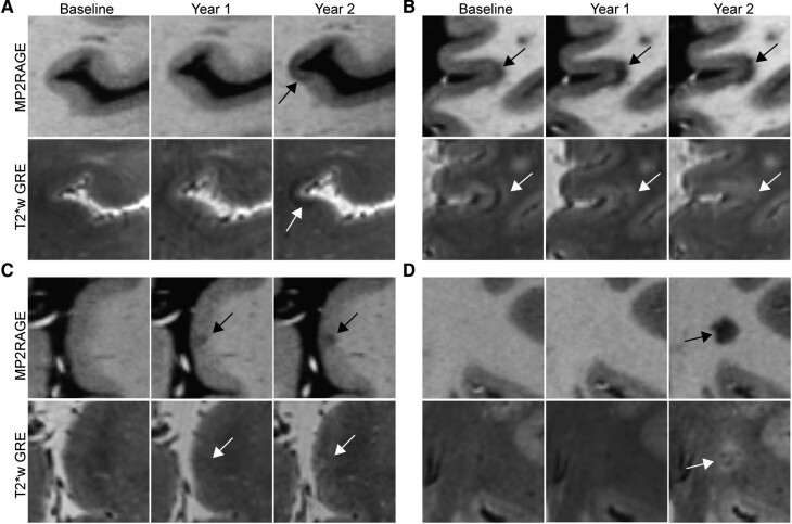
Appearance of new cortical lesions differs from the appearance of chronic cortical lesions. (A) A subpial lesion that was new on the Year 2 MRI appears hypointense on T2*w GRE. (B) A leukocortical lesion that was present at baseline is initially hyperintense on T2*w GRE with a band of hypointensity at the cortex-white matter junction. This lesion expands over time and becomes more hypointense on T1w MP2RAGE, while at the same time, the hypointense band on T2*w GRE disappears. (C) A subpial lesion that was new on Year 1 MRI initially appears isointense on T2*w GRE and then becomes more hyperintense at Year 2. (D) A white matter lesion in the same individual as (C) was new on Year 2 MRI and is hyperintense on T2*w GRE. Lesions are denoted with arrows. MP2RAGE, magnetization prepared 2 rapid acquisition gradient echo; T2*w GRE, T2*-weighted gradient-recalled echo.

