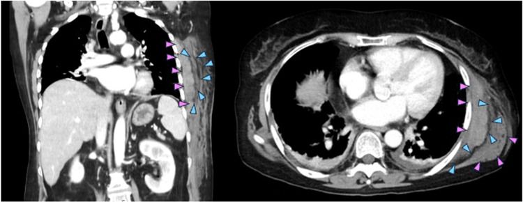
Figure 3. Contrast-enhanced computed tomography of the left lateral chest wall.
Contrast-enhanced computed tomography revealed a subcutaneous mass in the left lateral chest wall. This mass was characterized by fluid collection along with fascia of left serratus anterior (blue arrowheads) and swelling of left serratus anterior (pink arrowheads).

