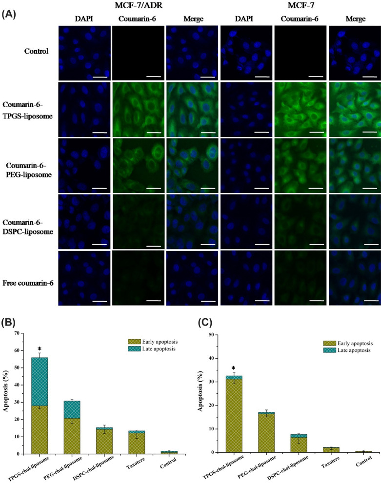
Fig. 7.
DTX-loaded NPs within liposomes. A Assessment of liposomal penetration into MCF-7 and MCF-7/ADR cells using confocal laser scanning microscopy (CLSM) after a 2-h exposure to different liposomal formulations, including free coumarin-6, TPGS-coumarin-6 liposome, PEG-coumarin-6 liposome, and DSPC-coumarin-6 liposome. The scale bar is set at 50 µm. The capacity of the various liposomal configurations to induce apoptosis in B MCF-7 cells and C MCF-7/ADR cells, with statistical significance denoted by *P < 0.05 when compared to TPGS-chol-liposome (n = 3)
(Reprinted with permission from [89])

