Fig. 1.
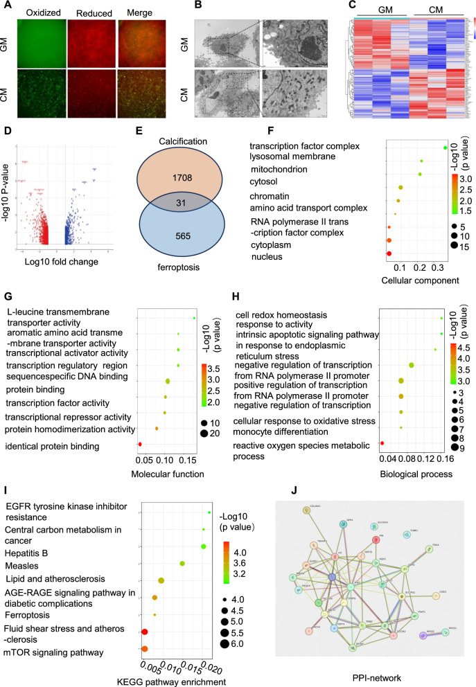
Ferroptosis contributes to vascular calcification dynamics. VSMCs were treated with high phosphate and calcium levels to induce VSMC calcification in vitro, and C11-BODIPY 581/591 was used to analyse fluorescent cytosolic and lipid ROS (A). Ultrastructural changes in muscle cells were examined using electron microscopy (B). Heatmap illustrating Differentially Expressed Genes (DEGs) between control and calcification groups in the GSE 211722 dataset (C). Volcano plots showing DEGs (D). Venn diagram illustrating overlapping DEGs identified in the two datasets (E). Gene Ontology enrichment analysis (biological processes, molecular functions, and cellular components) was conducted for the hub genes (F-H). Pathway analysis based on the Kyoto Encyclopaedia of Genes and Genomes was performed for hub genes (I). A Protein–Protein Interaction (PPI) network analysis was conducted for hub genes (J)

