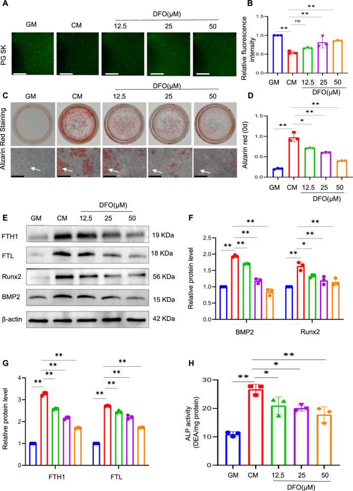
Fig. 3.
Inhibition of iron accumulation attenuates calcium/phosphate-induced VSMC calcification. Phen Green SK was used to evaluate intracellular Fe.2+ in VSMCs. Scale bar: 50 µm (A and B). After being treated for 14 days with GM or CM, with or without different concentrations of DFO (12.5, 25, 50 µM), calcification in VSMCs was detected by alizarin red staining and quantitative analysis. White arrow head indicates the calcified deposits. Scale bar: 100 µm (C and D). protein expression of osteogenic markers Runx2, BMP2, FTL, and FTH1 in VSMCs treated with GM or CM, with or without different concentrations of DFO (12.5, 25, 50 µM), was analysed by western blot and quantified by densitometry *P < 0.05, **P < 0.01 (E‒G). ALP activity in VSMCs was measured; *P < 0.05, **P < 0.01 (H)

