Fig. 5.
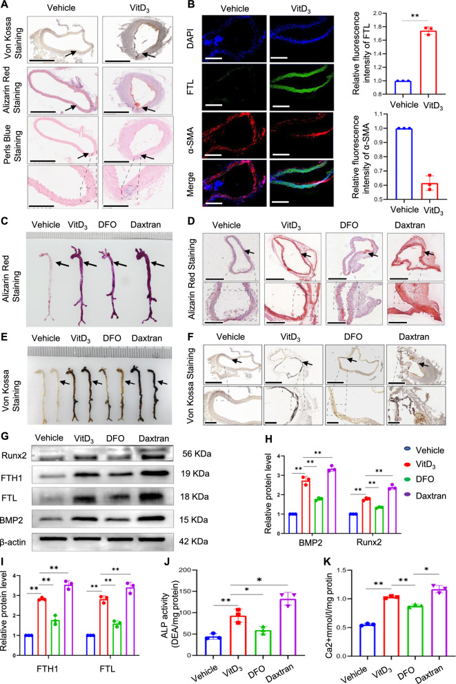
Iron overload is positively correlated with vascular calcification in mice. Mice vascular calcification was induced by injecting VitD3. Aortic sections were used to determine calcification by alizarin red and von Kossa staining; iron accumulation in aortic tissue was detected by Perl's blue staining. Black arrow head indicates the medial layer of artery. Scale bar: 300 µm (A). α-SMA and FTL in mouse arterial sections were assessed using immunofluorescence. Scale bar: 20 µm (B). The whole artery and thoracic aorta (indicated by black arrows) cross sections were used to compare calcification by using alizarin red S (C and D) and Von Kossa staining. Scale bar: 300 µm (E and F). Total aortic proteins were used to determine the expression of Runx2, BMP2, FTL, and FTH1 by western blotting (*P < 0.05, **P < 0.01, G–I). ALP activity in mouse aortas was measured (J). Mineral deposition in the entire artery was measured using calcium content assay (K)

