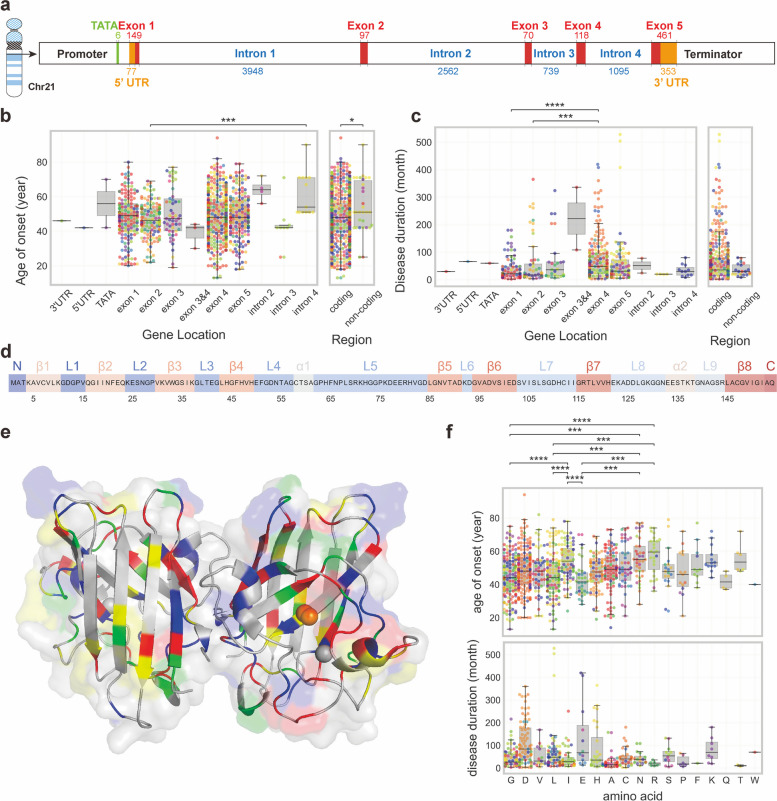
Fig. 3.
Molecular alterations of SOD1 variants. a Location of SOD1 gene (Accession No: NC_000021). b Age of onset distribution among patients with different genetic locations of SOD1 variants. c Disease durations of patients with different genetic location of SOD1 variants. d Secondary structure of SOD1 protein. The secondary structural elements, including alpha-helices and beta-strands, were identified using the PyMOL software (version 2.5.0) based on the 3D crystal structure of the human SOD1 protein (PDB id: 2V0A). e The 3D crystal structure of the human SOD1 protein from the Protein Data Bank (PDB id: 2V0A). Codon colors represent the associations of the variants with the mean disease duration and the mean age of onset. Green represents disease duration longer than 56.17 months (the mean disease duration of the total SOD1-related ALS patients) and the age of onset older than 48.51 years (the mean age of onset of the total SOD1-related ALS patients), blue indicates longer duration and younger age, yellow signifies shorter duration and older age, and red indicates shorter duration and younger age. (f) Age of onset and disease duration of patients with different amino acid sites of variant. Significance was analyzed using Kruskal–Wallis test and P-values were adjusted to account for multiple comparisons using Bonferroni correction. *P ≤ 0.05, ** P ≤ 0.01, *** P ≤ 0.001, ****P ≤ 0.0001

