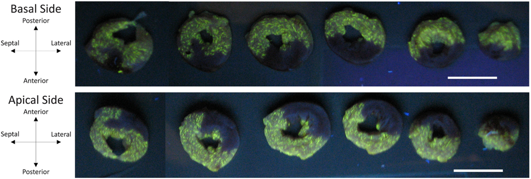
Fig. 1.
Short-axis sections of a representative left ventricle under ultraviolet light following arrest by retrograde perfusion with cold 2,3-butanedione monoxime phosphate-buffered saline containing fluorescent microsphere enables visualization of infarct region. Coronary artery ligation prevented microspheres from reaching infarcted myocardium (dark regions). Scale bars are both 10 mm in length.

