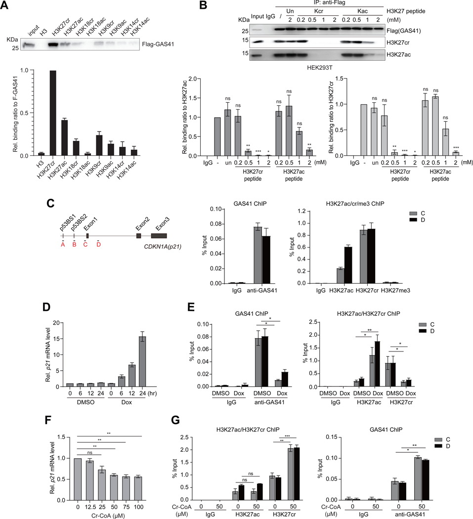
Figure 2. GAS41 regulates transcriptional repression of p21 mediated by H3K27cr.
(A) Western blots analysis and quantitation of ectopically expressed Flag-GAS41 in HCT116 cells binding to a set of biotinylated histone H3 peptides derived from H3 (residues 1–25, biotin-ARTKQTARKSTGGKAPRKQLATKAA), H3K27cr/ac (residues 21–33, biotin-ATKAAR-Kcr/Kac-S-APATG), H3K18cr/ac (residues 12–24, biotin-GGKAPR-Kcr/Kac-QLATKA), H3K9cr/ac (residues 3–15, biotin-TKQTAR-Kcr/Kac-SSGGKA), and H3K14cr/ac (residues 1–25, biotin-ARTKQTARKS-TGG-Kcr/Kac-APRKQLATKAA). n=3.
(B) Western blot analysis and quantitation of Flag-GAS41 binding to nucleosomal H3K27cr or H3K27ac in the lysate of HEK293T cells transiently transfected with Flag-GAS41 in the presence of unmodified histone H3K27, H3K27cr or H3K27ac peptide at concentrations as indicated. n=3.
(C) Scheme illustrating primer positions at p21 gene locus used in the study. Right, ChIP-qPCR analyses of endogenous GAS41(left), and histone H3K27cr, H3K27ac, or H3K27me3 (right) enrichment at p21 locus in HEK293T cells. n=3.
(D) mRNA transcript levels of p21 in HEK293T cells treated with Dox for 0, 6, 12, and 24 hours, respectively, n=3.
(E) ChIP-qPCR analyses showing changes of endogenous GAS41 (left), and H3K27ac or H3K27cr (right) enrichment at p21 locus in HEK293T cells treated with Dox for 24 hours, n=3.
(F) qPCR analysis of p21 expression in HEK293T cells treated with crotonyl-CoA at concentrations as indicated for 24 hours. n=3.
(G) ChIP-qPCR analysis of H3K27ac, H3K27cr (left), and endogenous GAS41 (right) enrichment at p21 locus in HEK293T cells with 24-hour crotonyl-CoA (50 μM) treatment. n=3.

