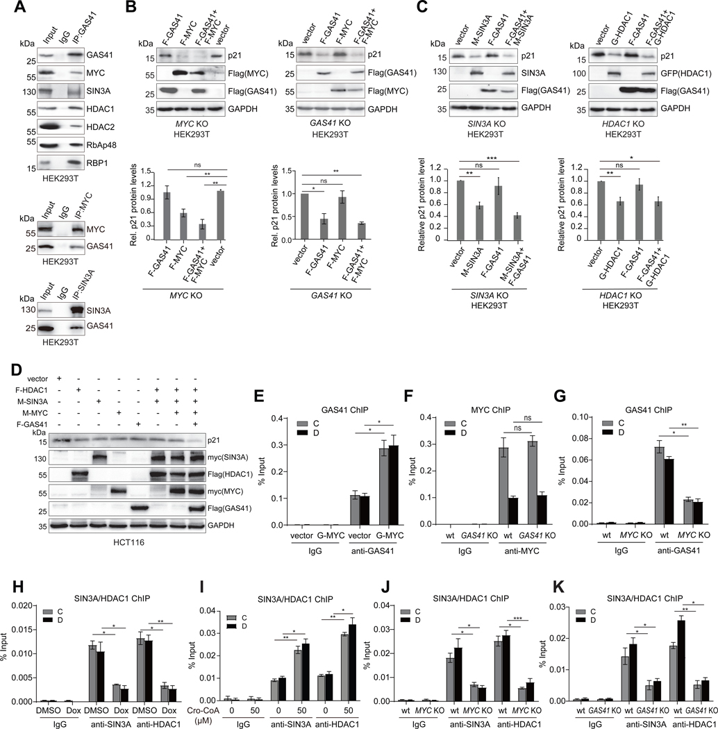
Figure 3. GAS41 works with MYC and SIN3A/HDAC1 co-repressors for p21 repression.
(A) Co-immunoprecipitation (Co-IP) of endogenous GAS41 and immunoblotting (IB) with specific antibodies assessing GAS41 association with MYC, SIN3A, HDAC1/2, RbAP48 and RBP1 in HEK293T cells (upper). Lower, reverse Co-IP showing endogenous MYC and SIN3A interactions with GAS41, respectively, in HEK293T cells.
(B) Western blot analysis and quantification of p21 expression in HEK293T MYC KO (left) and GAS41 KO (right) cells co-transfected with F-GAS41 and F-MYC, n=6 for MYC KO, n=3 for GAS41 KO.
(C) Western blot analysis and quantification of p21 expression in HEK293T SIN3A KO cells co-transfected with F-GAS41 and M-SIN3A (left), and HDAC1 KO cells co-transfected with F-GAS41 and G-HDAC1 (right). n=5 for SIN3A KO, and n=6 for HDAC1 KO.
(D) Western blot analysis of p21 expression in HCT116 cells co-transfected with F-HDAC1, M-SIN3A, M-MYC, and F-GAS41.
(E) ChIP-qPCR analysis of GAS41 enrichment at p21 locus in HEK293T cells transiently co-transfected with GFP empty vector, or GFP-MYC. n=3.
(F) ChIP-qPCR analysis of endogenous MYC enrichment at p21 locus in HEK293T wt and GAS41 KO cells. n=3.
(G) ChIP-qPCR analysis of GAS41 enrichment at p21 locus in HEK293T wt and MYC KO cells. n=3.
(H) ChIP-qPCR analysis of SIN3A and HDAC1 enrichment at p21 locus in HEK293T cells before and after 24-hour Dox treatment. n=3.
(I) ChIP-qPCR analysis of SIN3A/HDAC1 enrichment at p21 locus in HEK293T cells with 24-hour crotonyl-CoA (50 μM) treatment. n=3
(J) ChIP-qPCR analysis of SIN3A and HDAC1 enrichment at p21 locus in HEK293T wt and MYC KO cells. n=3.
(K)ChIP-qPCR analysis of SIN3A and HDAC enrichment at p21 locus in HEK293T wt and GAS41 KO cells. n=3. F-GAS41 stands for Flag-GAS41, F-MYC for Flag-MYC, F-HDAC1 for Flag-HDAC1, G-HDAC1 for GFP-HDAC1, M-SIN3A for myc-SIN3A, and M-MYC for myc-MYC.

