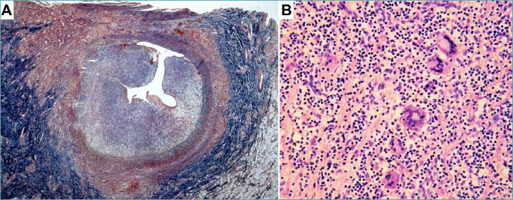
Figure 3.

Coronary artery vasculitis in Takayasu’s disease. A 14-year-old girl was rescued after a sudden cardiac arrest. Aortography showed mild aortic dilation with aortic valve insufficiency and sub-occlusion of coronary ostia. The girl died and the autopsy revealed massive myocardial infarction with thickening of the ascending aorta, aortic arch, pulmonary arteries, left external carotid artery, and coronary ostia (modified from 82). (A) The left anterior descending coronary artery shows severe luminal narrowing because of concentric intimal hyperplasia. The tunica media appears destroyed and the adventitia is fibrotic. (B) The involved segments reveal necrotizing granulomatous inflammation with giant cells, consistent with Takayasu’s arteritis. (A) Masson trichrome stain; (B) Hematoxylin-eosin stain.
