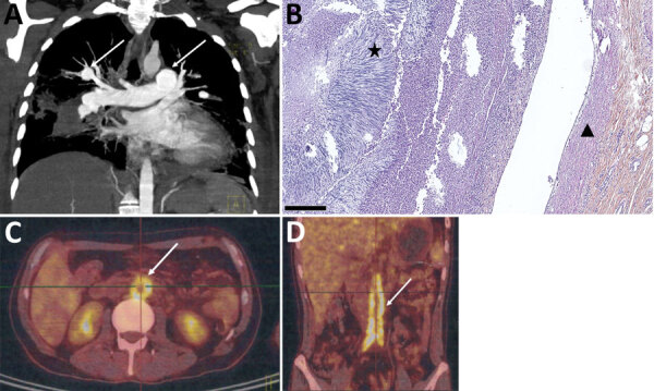
Figure 2.

Imaging of pulmonary arteritis and abdominal aortitis for case-patients 2 and 3 in study of unexpected vascular locations of Scedosporium spp. and Lomentospora prolificans fungal infections, France. A) Thoracic computed tomography scan for case-patient 2. Arrows indicate left pulmonary artery and right lobar pulmonary artery mycotic aneurysms. B) Hematoxylin-eosin-saffron stain of lung tissue from postmortem analysis of case-patient 2. Triangle indicates bronchial artery wall; star indicates bronchial artery thrombus consisting of radially-disposed multiple septate hyphae. Scale bar indicates 250 μm. C, D) Positron emission tomography-computed tomography scans of case-patient 3. C) Arrow indicates intense abdominal aorta hypermetabolism. D) Arrow indicates abdominal aorta and primitive iliac artery hypermetabolisms. Data are from the Scedosporiosis/lomentosporiosis Observational Study (12).
