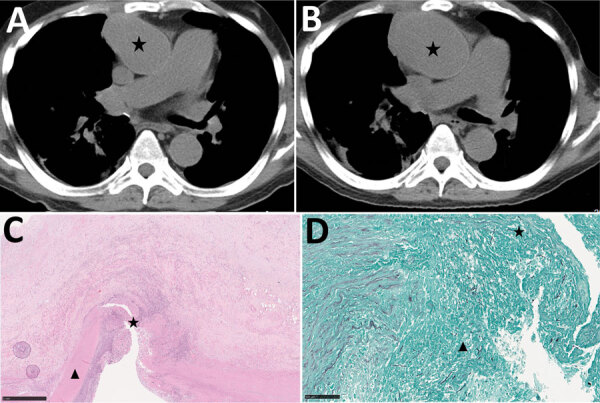
Figure 5.

Radiologic and histopathologic analysis of thoracic aortitis in case-patient 6 in study of unexpected vascular locations of Scedosporium spp. and Lomentospora prolificans fungal infections, France. A) Thoracic computed tomography scan showing sacciform aneurysm of ascending aorta (star). B) Thoracic computed tomography scan 21 days later showing the rapidly growing aneurysm (star). C) Hematoxylin-eosin-saffron stain of thoracic aorta section. Star indicates thoracic aorta wall dissection. Triangle indicates tunica media. Scale bar indicates 1 mm. D) Grocott-Gomori methenamine silver stain of thoracic aorta section. Star indicates septate fungal hyphae invading the thoracic aorta tunica media. Triangle indicates thrombus containing septate fungal hyphae. Scale bar indicates 100 μm. Data are from the Scedosporiosis/lomentosporiosis Observational Study (12).
