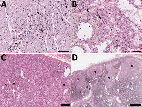
Figure 3.

Histology section of thawed, formalin-fixed tissues from harbor seals (Phoca vitulina) infected by highly pathogenic avian influenza A(H5N1) virus in the St. Lawrence Estuary, Quebec, Canada, 2022. Hematoxylin phloxine saffron stain. A) Brain tissue from a young (<1 year old) female seal. The Virchow-Robin space around a vessel (V) is infiltrated by numerous layers of polymorphonuclear cells. Several neurons have a condensed hyperacidophilic cytoplasm indicative of necrosis and are often associated with satellitosis (arrows). A focally extensive infiltration of the neuropil by neutrophils and glial cells is also present. Scale bar indicates 100 µm. B) Lung from a young (<1 year old) female seal. The alveolar (A) and vascular (arrow) lumens contain numerous, often degenerate, polymorphonuclear cells. The alveolar walls are infiltrated by numerous inflammatory cells composed of neutrophils and mononuclear cells. The epithelial cells bordering the small bronchi are often necrotic. Scale bar indicates 100 µm. C) Adrenal gland of an adult female seal. Multifocal foci of necrosis are present in the cortical zone (N). Scale bar indicates 300 µm. D) Lymph node from an adult female seal. Marked multifocal to coalescing necrosis of the lymphoid tissues in the cortical region are noted (N). Scale bar indicates 1 mm.
