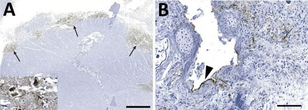
Figure 4.

Detection of influenza virus antigen by immunohistochemistry in brain (A) and lung (B) of harbor seals (Phoca vitulina) infected by highly pathogenic avian influenza A(H5N1) virus, St. Lawrence Estuary, Quebec, Canada, 2022. A) Brain tissue. Multifocal areas of intense immunostaining (arrows) with staining of all structures are seen in the affected area, including neurons and neuropil (inset). Scale bar indicates 2 mm. B) Lung tissue. Positive immunostaining can be observed within alveolar septae (arrow) and in bronchiolar epithelial cells (arrowhead). Scale bar indicates 80 µm.
