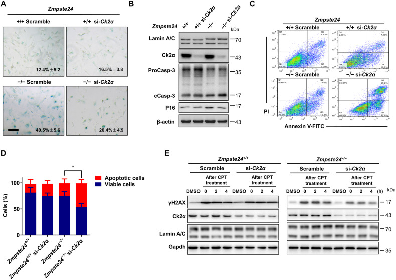
Fig. 4. TBB-induced apoptosis in Zmpste24-deficient cells via inhibition of CK2.
A Senescence-associated β-galactosidase staining in Ck2α or scramble siRNA treated Zmpste24+/+ and Zmpste24−/− MEFs at passage 6 (scale bar, 200 μm). B Representative immunoblots showing the levels of proCaspase-3 (ProCasp-3), cleaved caspase-3 (cCasp-3) and P16 in Ck2α or scramble siRNA-treated Zmpste24−/− MEFs and wild-type controls. C Representative flow cytometric plots for assessing apoptosis in Ck2α or scramble siRNA-treated Zmpste24−/− MEFs and wild-type controls. D Quantitation of the percentage of viable (gate II: PI−/annexin V−) and apoptotic (gates III and IV: PI−/annexin V+ and PI+/annexin V+) cells in (C); chart plots mean ± SD from independent triplicate experiments. E Representative immunoblots showing protein levels of γH2AX at indicated time points after 4 μM camptothecin (CPT) for 1 h to induce DNA damage or vehicle DMSO control in siRNA-Ck2α transfected in Zmpste24−/− MEFs and wild-type controls treated with 75 μM TBB for 6 h at the indicated time (after CPT treatment 0, 2, 4 h). At least three independent experiments were performed.

