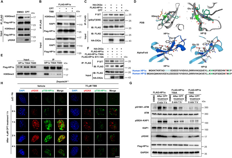
Fig. 5. TBB affects the phosphorylation of HP1α T50 by CK2α, leading to an impaired DNA damage response in Zmpste24-deficient MEFs.
A Immunoblots showing H3K9me3 protein in anti-Flag immunoprecipitates in HEK293 cells expressing FLAG-HP1α treated with 4 μM CPT for 1 h. B Representative immunoblots from at least three independent experiments showing HEK293 cells expressing FLAG-HP1α treated with 4 μM camptothecin (CPT) for 1 h to induce DNA damage or vehicle DMSO control pretreated with 75 μM TBB or mock DMSO (control) for 6 h. C Immunoblots showing p-S/T or phospho-CK2 Substrate [(pS/pT)DXE] of FLAG-HP1α transfected HA-CK2α and FLAG-HP1α treated with 4 μM camptothecin (CPT) for 1 h to induce DNA damage or vehicle DMSO control by using an anti-FLAG antibody immunoprecipitates in HKE293 cells. D A model for the interaction of the human HP1α (left) and HP1-β (right) chromodomain bound to methylated K9 of histone H3 (K9me3), based on the PDB protein database and AlphaFold. A network of hydrogen bonds between the side chains of Glu 52, Thr 50 and Leu 39 of the human HP1-α chromodomain; Glu 53, Thr 51 and Leu40 of the human HP1-β chromodomain. E Immunoprecipitation showing the binding capacity between H3K9me3 and various FLAG-HP1α mutants. F Immunoblots showing p-S/T of FLAG-HP1α or FLAG-HP1α mutants transfected HA-CK2α by using an anti-FLAG antibody in HKE293 cells. G Representative immunoblots showing protein levels of ATM-Ser1981, KAP1-Ser824 and γH2AX in FLAG-HP1α and T50A or T50D mutants transfected in HEK293 cells. Note that the response to DNA repair is less sensitive in FLAG-HP1α T50A or T50D mutants because of lower ATM-Ser1981, KAP1-Ser824 and γH2AX expression. Cells were treated with 4 μM camptothecin (CPT) for 1 h or vehicle DMSO and then analyzed by western blotting. Data represent independent triplicate experiments. H Representative immunofluorescence confocal microscopy images of γ-H2AX and pT50-HP1α immunofoci recruitment for each condition in response to 4 μM CPT-induced DNA damage in TBB treated Zmpste24–/– MEFs. TBB inhibited the co-immunofoci of pT50-HP1α and γ-H2AX at the indicated time (after CPT treatment 0, 1, 2, 4 h). Scale bar, 10 µm.

