
FIGURE 2.
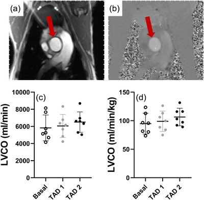
Representative anatomical‐magnitude (a) and phase (b) images of the maternal ascending aorta utilized for the determination of maternal left ventricular cardiac output (LVCO) unindexed (c) and indexed (d) to maternal weight across basal (open circles), TAD 1 (grey filled circles) and TAD 2 (black filled circles) periods. Data presented as individual data points with means ± SD superimposed. Data analysed by a repeated measures one‐way ANOVA with Bonferroni's correction for multiple comparisons.